THE BARN RAT
  • Learning Center
    • Anatomy
    • Barn Tours
    • Culture
    • Equipment
    • Grooming
    • Product Reviews
    • Riding
    • Transformations
    • Videos
  • Career Center
    • Farrier School
    • Vet School
  • Book Club
  • Library
    • Magazines
    • Movies
  • Blog
  • Shop
  • Learning Center
    • Anatomy
    • Barn Tours
    • Culture
    • Equipment
    • Grooming
    • Product Reviews
    • Riding
    • Transformations
    • Videos
  • Career Center
    • Farrier School
    • Vet School
  • Book Club
  • Library
    • Magazines
    • Movies
  • Blog
  • Shop

About: Bone Anatomy of a Horse's Front Leg

4/25/2018

1 Comment

 
Picture
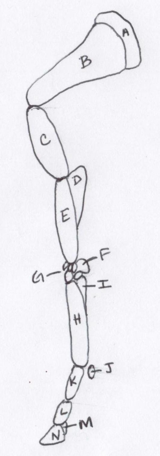
   In this next part of our anatomy series we will focus on the bones of the front limb. The image below is not accurately to scale but it does show the general areas in which these bones live.
Picture

Picture
A - Suprascapular Cartilage

B - Scapula: The "shoulder blade"; Transmits forces and motion of the front limb; Attachment of muscles and connective tissue that support cranial portion of the body; Flat in shape

C - Humerus: Attachment of major muscles that move upper and lower limb; "Point of Shoulder"

D - Ulna: Fused to the Radius to prevent supination (the outward rotation of the limb); "Point of Elbow"

E - Radius: optimizes cranial (closer to the head or top) and caudal (closer to the feet or bottom) muscles

F - Accessory Carpal: always lateral (or on the outside); the retinaculum attached as shown below to restrict other structures (tendons) to stay in place
Picture
G - Carpus: 7 bones comparable to a human wrist; provide shock absorption and flexion

H - Metacarpal III (Cannon Bone): comparable to the long bones in a human hand

I - Metacarpal IV (Splint Bone): 2 splint bones on each leg; creates a channel for the tendons to settle within; provides support to the cannon bone

J - Proximal Sesamoid: embeded in the suspensory ligament; prevent overextension of the fetlock joint

K - Proximal Phalanx (Long Pastern Bone): comparable to the first bone in a human finger

L - Intermediate Phalanx (Short Pastern Bone): similar to the middle medium sized bone in a human finger

M - Distal Sesamoid (Navicular Bone): helps to stabilize the lower joints and the hoof; many ligaments in this area

N - Distal Phalanx (Coffin Bone): similar to the last small bone in a human finger where the fingernail grows
1 Comment
Iowa Granny Hookup link
11/11/2022 12:31:34 am

This is a great posst thanks

Reply


Leave a Reply.

    Picture

    Instagram


    Categories

    All
    About
    Anatomy
    Barn Tours
    Book Club
    Book Review
    Career Panel
    Clipping
    College
    Equipment
    Etiquette
    Grooming
    How To
    Interviews
    Product Review
    Riding
    Road To RRP 2019
    Show Ready
    Transformations
    Videos


    Picture
    * indicates required

    Sponsors

    Cantera Equestrian
    Cantera Equestrian

    Picture
    Trafalgar Square Books
    Interested in sponsoring The Barn Rat? Be sure to contact us here!
© COPYRIGHT 2022. ALL RIGHTS RESERVED.